WhatsApp

Ultrasound

Unveiling Your Inner World

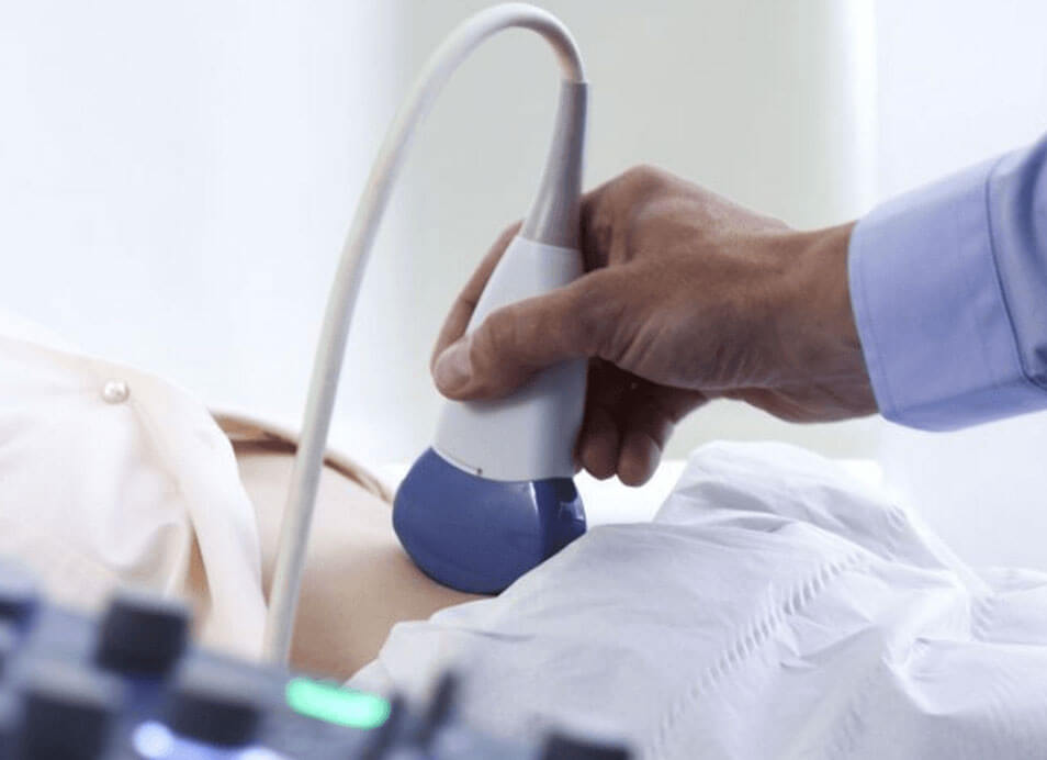
At Ketkar Maternity Hospital and Research Centre, we understand the power of visualization. Ultrasound, also known as sonography, serves as a window into your body, allowing us to capture detailed images of your internal organs and tissues. This non-invasive and painless imaging technique uses sound waves to create real-time pictures, providing valuable insights into your health and aiding in accurate diagnosis and treatment planning.

Accurate Diagnosis, Personalized Treatment

At Ketkar, we pride ourselves on utilizing the latest ultrasound technology and experienced sonographers to deliver accurate and reliable diagnostic imaging. Our multidisciplinary team collaborates to interpret the results, ensuring a personalized treatment plan tailored to your specific needs.

Comfort and Convenience

We strive to create a comfortable and stress-free environment for all our patients. Our ultrasound suites are equipped with advanced imaging technology and staffed by compassionate professionals who prioritize your well-being. We also offer flexible appointment scheduling to accommodate your busy lifestyle.


Common Questions

If you could not find an answer to your query, please whatsapp us.

An ultrasound, also known as a sonogram, is a diagnostic imaging technique that uses high-frequency sound waves to create images of the inside of the body. It is commonly used in pregnancy to monitor the development of the fetus and in diagnosing various medical conditions.

During an ultrasound, a device called a transducer emits sound waves that bounce off tissues and organs. These echoes are then captured by the transducer and converted into images by a computer. The images provide real-time visual information about the structures being examined.

Yes, ultrasound is considered safe and non-invasive. It does not use ionizing radiation, like X-rays, and has been widely used in medical practice for decades without known harmful effects when performed properly.

Ultrasound is used in pregnancy to monitor the development and health of the fetus, check for multiple pregnancies, estimate the due date, assess the position of the placenta, detect fetal abnormalities, and evaluate amniotic fluid levels. It helps ensure both maternal and fetal well-being.

The first ultrasound, often called a dating scan, is typically performed between 8 and 14 weeks of pregnancy. It confirms the pregnancy, estimates the gestational age, checks for a heartbeat, and identifies the number of fetuses.

A transvaginal ultrasound is an internal ultrasound where a transducer is inserted into the vagina to provide clearer images of the pelvic organs, including the uterus and ovaries. It is commonly used in early pregnancy and to investigate gynecological conditions.
RAISE A QUERY

Contact Us

Get In Touch

Address

Ketkar Maternity Hospital & Research Institute

Temple Bazar Road, Opp. St. Xavier Church, Behind Moonlight Photo Studio, Sitabuldi, Nagpur, Maharashtra 440012

Phone No.

Expert Tips from Our Consultant